Ptosis is an abnormal low position of the
upper eyelid which may be congenital or acquired. It is a common problem and is
found in all age groups. Primary congenital ptosis is present at birth and
tends to be non progressive. It may be bilateral, isolated, or part of an
associated syndrome. There is harmony between its severity and levator
function. It is often due to the poor development of the levator muscle or its
replacement by fibrosis, fat, or areolar tissue1.
Amblyopia is rare in congenital ptosis unless it is associated with severe
unilateral ptosis, anisome-tropia, or strabismus2. Anatomically
ptosis may be classified as neurogenic (third nerve palsy, Horner syndrome, and
Marcus Gunn Jaw-winking syndrome), myogenic (myasthenia gravis, myotonic
dystrophy, ocular myopathy, simple congenital, or blepharophi-mosis syndrome),
aponeurotic (involutional, postoperative), and mechanical (dermatochalasis,
tumors, edema, anterior orbital lesions, and scarring)3.
To classify a ptosis into one of these categories, a thorough medical
history and physical examination must be performed and certain tests may be
necessary. There are three classic al surgical procedures for the treatment of
Ptosis; frontalis suspension, levator resection and Muller muscle-conjunctival
resection. Frontalis sling is considered as only option for poor levator
function Ptosis. We conducted this study to observe the usefullness of levator
resection as primary surgical procedure in all congenital ptosis patients with
poor upper lid excursion.
MATERIAL AND
METHODS
Study Design: It was a single centre, prospective, interventional case series. The study was
carried out in duration of more than two calendar years starting from March
2008 to November 2010.
Setting: The study was conducted at the
Department of Ophthalmology, a tertiary eye care and teaching facility, at
Bahawal Victoria Hospital, Bahawalpur, affliated with Quaid-e-Azam Medical
College Bahawalpur.
Sample: We treated
56 eyes of 50 patients. All had poor levator function, good Bell's phenomenon,
normal pupil size and reaction to light and normal corneal sensitivity. Males were 36 and 14 were female. Age range was 04-32 years
(Mean: 14 years).
Diagnosis was based on history, old photographs,
and routine ophthalmic examination. Oculoplastic examination specific to ptosis
was performed by the operating surgeon, this included, vertical palpebral
fissure height, Marginal reflex distance (MRD), levator excursion, lid crease
height, Bells phenomenon and ocular motility. All patients included were
diagnosed as congenital ptosis. It also included
checking head position, chin elevation, brow position, and brow action in
attempted up gaze. All the patients had detailed systemic evaluation to rule out
secondary causes of the ptosis.
Exclusion criteria were, absent Bell’s phenomenon,
disturbed or absent corneal sensitivity and dry eyes. Surgery was performed by single surgeon (RRQ). All patients were explained about the procedure and informed consent obtained.
Definitions
Excellent: 0 and +/- 0.5 mm
and complete lid closure.
Good: +0.5 mm and +1.00 mm and complete lid closure.
Fair: + 1.00 mm and + 1.5 mm and complete lid closure.
Poor: greater than +
1.5 mm.
Technique of Surgical
Intervention: Levator Resection was
carried out through skin approach (Blascovics technique). All patients were subjected to general anesthesia. After preparing and
draping, an incision was marked at a level symmetric with the opposite eyelid
usually 8-10 mm above the lid margin. A cut was made along the marked line
using #15 scalpel blades. A blunt dissection was carried out towards lid margin
to expose tarsal plate for re-attachment of levator at the end of the surgery.
The post orbicular facial plane was entered and orbital septum was exposed and
confirmed by applying inward pressure at lower part of globe and pre
aponeurotic fat popped up under septum. The septum was incised with sharp
scissors and the attachments between the septum and aponeurosis were separated
to prevent postoperative lagophthalmos. The aponeurosis and Whitnall's ligament
were revealed by brushing the pre aponeurotic fat pockets upward. This was
followed by disinsertion of the aponeurosis from the tarsus. Carrying blunt
dissection, the muscle was dissected all the way to the Whitnall's ligament. A
6.0 vicryl was passed through partial thickness of the tarsus, 3 mm from its
upper border and above the central pupil posterior to the aponeurosis and
retrieved through the Whitnall's. Two additional sutures were added between the
tarsus and Whitnall’s and placed medially and laterally. The three sutures were
adjusted as needed. Finally, the skin incision was closed with running 6.0
vicryl sutures.
Complications: Major per operative complications faced were loss of proper facial
plane, hemorrhage while separating aponeurosis from conjunctiva and button
holes in conjunctiva.
Follow-ups: Patients had a follow-up on day one, at 4 weeks, 6 months and then last
follow up at 2 years.
RESULTS
Goal was to adequately elevate the lid while minimizing the risk of
lagophthalmos and exposure keratopathy/ulceration. In majority (85.65%) results obtained were good to excellent (Table 1)
with a well-defined symmetry in lid height and shape (Fig 1-3). In four (7.14%)
cases, results were cosmetically acceptable and patients were satisfied
although graded as fair, however residual ptosis occurred in four cases (7.14%)
and required further surgical procedure at a later date. Reoperation was
uncomplicated and final outcome was successful. The significant postoperative
complications were over correction in
one patient which was not significant to warrant reoperation.
One female patient had forniceal prolapsed (Fig 4) which was sutured and
two patients had suture related granuloma, treated with antibiotics, which
did not influence the final outcome.
DISCUSSION
Embryologically,
most of the connective tissue of upper lid is derived from mesenchyme15-17,21. The orbital septum is
derived from mesenchyme of second arch15. Suborbicularis fibro adipose
tissue consists of multiple fibrous septa that merge posteriorly with the
orbital septum and give orbital septum a multilayered quality, augmenting the
contour of superior sulcus6,22. Simple congenital ptosis is thought
to be the result of
Pre-op
Post-op
Fig 1:
Pre-op
Post-op
Post-op
lid closure
Fig 2:
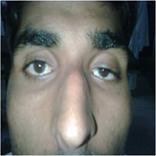
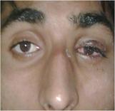
Fig 3: Pre-op
Post-op
Fig 4: Suture to
forniceal prolapse
Table 1:
|
Outcome |
No. of patients n (%) |
|
Excellent |
38 (67.8) |
|
Good |
10 (17.85) |
|
Fair |
04 (7.14) |
|
Poor |
04 (7.14) |
|
Total |
56 (100) |
developmental
dystrophy of levator muscle. Normal muscle fibres are replaced by fibrous
connective tissue without contractile properties. Ptosis is more marked in an
up gaze and the upper lid is relatively retracted in a down gaze16.
Ptosis can have a
marked impact on a patient's functional status9 and lead to poor
visual development in childhood with its damaging social and financial
consequences in later life2. The goal of ptosis surgery was once
described as one with elusive perfect result10. Ptosis surgery in
paediatric patients differed from adult surgery in that predictability of lid
height in later group could be enhanced by using local anaesthesia or
adjustable sutures11,12. As there were no authentic published data
regarding time taken to reach final lid height stability in primary congenital
ptosis patients, we chose a maximum follow-up of 2 years as a stable end point.
In ptosis surgery,
a good cosmetic outcome is very important, this holds true for congenital
myogenic ptosis as well. More than 100 techniques for the treatment of ptosis
have been reported4-6. This means ptosis is difficult to treat, as
the postoperative eyelid position may be unpredictable20. Different surgical techniques have been laid
out for the management of primary congenital ptosis. This depends upon severity
of ptosis, laterality, and levator function. The surgical approach may include
posterior resection for mild ptosis with normal levator function or levator
aponeurosis resection for moderate-to-poor levator function and frontalis
suspension for bilateral ptosis with poor to absent levator
function8. In our patients, levator aponeurosis resection has
given the best results with excellent patient satisfaction despite the fact
that the levator function was extremely poor (<4 mm).
Although it has
been reported that extra-large levator resection may lead to lagophthalmos,
none of our patients has experienced this complication. The lagophthalmos may
not be a problem as it depends on the orbicularis tone and function. Every
ptosis surgery has goals such as controlled height, contour, lid crease, lash
position, and symmetry. We found that our patients achieved almost all such
targets.
In ptosis surgery, use of adjustable suture technique is popular in
adults but not well tolerated in children. It is therefore important to
consider an approach that gives good ptosis correction with cosmetically
acceptable upper lid skin crease19. The ideal procedures
in ptosis surgery are those that disturb normal anatomy the least and also
allow for good results17. In this study an anterior approach was
selected, thus avoiding conjunctiva, lacrimal gland and tarsus. In all cases,
after incising skin, blunt dissection in a proper facial plane was carried out
to reveal septum. Incising septum gave the hold of aponeurosis and separation
of it from underlying conjunctiva is critical to avoid bleeding from peripheral
vascular plexus and saving conjunctiva from button holling. Finally, muscle is
attached to tarsus with 6-0 vicryl suture and skin is closed with the same type
of suture. This technique appears to enhance the overall cosmetic outcome.
CONCLUSION
In this series we
treated 56 eyes of 50 patients with primary congenital ptosis and poor levator
function with levator aponeurosis resection. All the patients achieved the desired result without any complications. Although
recent findings have shown the frontalis suspension technique is a commonly
performed surgical correction of congenital ptosis, used widely in the repair
of Ptosis with poor levator function, we recommend that levator resection
procedure to be considered as primary procedure for the correction of
congenital ptosis with very poor levator function.
Author’s affiliation
Dr.
Rao Muhammad Rashad Qamar
Associate Professor of
Ophthalmology
QAMC/BVH,
Dr. Muhammad Younis Tahir
Senior Registrar of
Ophthalmology
QAMC/BVH,
Dr.
Abid Latif
Senior Registrar of Ophthalmology
QAMC/BVH,
Dr. Ejaz Latif
Professor of
Ophthalmology
QAMC/BVH,
REFERENCE
1.
Baroody
M, Holds JB, Vick VL. Advances in the diagnosis and
treatment of ptosis. CurrOpin Ophthalmol. 2005; 16: 351-5.
2.
Anderson
RL, Baumgarter SA. Amblyopia in ptosis. Arch
Ophthalmol. 1980; 98: 1068-9.
3.
Finsterer
J. Aesthetic Plast Surg. Ptosis: Causes, presentation,
and management. 2003; 27: 193-204.
4.
Jones
LT. The anatomy of the upper eyelid ptosis
surgery. Am J Ophthalmol. 1964; 57: 943-59.
5.
Jones
LT, Quickert MH, Wobig IJ. Aponeurotic repair. Arch Ophthalmol.
1975; 8: 629-34.
6.
Mustardι
JC. Correction of ptosis by split level lid
resection. ClinPlas Surg.1978; 5: 533-5.
7.
Allard
FD, Durairaj VD. Current techniques in surgical
correction of congenital ptosis.
8.
9.
Federeci
TJ,
10.
Carraway
JH. Cosmetic and functional considerations in
ptosis surgery. The elusive "perfect" result. ClinPlas Surg. 1988;
15: 185-93.
11.
Lindberg
JV, Vasquez RJ, Ckao GM. Aponeurotic ptosis repair under
local anesthesia. Ophthalmology. 1988; 95: 1046-52.
12.
Collin
JR, O'Donnel BA. Adjustable sutures in eyelid
surgery for ptosis and lid retraction. Br J Ophthalmol. 1994; 78: 167-74.
13.
Complications of ptosis surgery and
their management. In: McCord C, editor. Eyelid surgery principles and techniques.
Chap. 11.
14.
15.
Bremond-Gignac
DS, Deplus S, Cussenot O. Anatomic study of orbital septum.
Surg Radiol Anat. 1994; 16: 121-4.
16.
Meyer
DR, Lindberg JV, Wobig JL. Anatomy of orbital septum and
associated eyelid connective tissues. Implications for ptosis surgery. Ophthal Plast
Reconstr Surg. 1991; 7: 104-13.
17.
Jones
LT. The anatomy of the upper eyelid and its
relationship to ptosis surgery. Am J Ophthalmol. 1964; 57: 943-59.
18.
Berke
RN,
19.
Collin
JRO, O’Donnell BA. Adjustable sutures in eyelid
surgery for ptosis and lid retraction. Br J Ophthalmol. 1994; 78: 167-74.
20.
Berke
RN,
21.
Wolff
E: Anatomy of eye and orbit. Philadelphia, Blackiston
Company. 1954: 153-209.
22.
23.
Baylis
HI,
24.
Mustarde
J, Callahan A, Jones L. Ophthalmic Plastic Surgery-
Up-to-Date.
25.
McElvanney
AM, Adhikary HP: Congenital ptosis, a good cosmetic
result with redefinition and suturing of the orbital septum. Eye. 1996; 10: 548-50.